Vein Occlusions
What are retinal vein occlusions?
A retinal vein occlusion occurs when one of the veins of the retina becomes blocked and cannot drain blood from the retina normally. This blockage leads to a backup of blood and fluid into the retina. This back up of blood can be seen in the retina as hemorrhages. The amount of swelling in the retina varies and depends on the degree of blockage of the retinal vein.
With retinal vein occlusion, weaker blood vessels may end up carrying more blood. They might start to leak, which causes the macula, the central part of retina, to swell and thicken. This is called macular edema, and it leads to blurry vision or vision loss. The swelling is treated so that the retina nerve cells do not drown.
With an occlusion, abnormal blood vessels can grow in your iris, the colored part of your eye). This may lead to a complication where the eye pressure rises and causes eye pain.
Two types of vein occlusions
Central Retinal Vein Occlusion (CRVO)

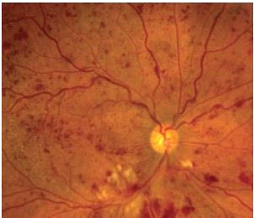
This is when your eye’s main vein is blocked, the central vein. This is located at the optic nerve. This causes hemorrhages to be diffusely scattered throughout the retina, and often affecting the central portion of the retina, the macula.
Branch Retinal Vein Occlusion (BRVO)

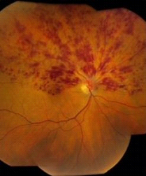
If the blockage involves only a branch of the central vein it is called a branch retinal vein occlusion. In these cases, hemorrhages and swelling are limited to the area of the retina that is drained by that particular vein. In the above picture the superior vein branch is blocked. The cause is usually from a diseased adjacent artery compressing the vein wall at a crossing point in the retina. The artery wall is likely diseased by arteriosclerosis, diabetes, hypertension or any combination
What causes retinal vein occlusion?
It is not known exactly what causes a central retinal vein occlusion. It may be due to several problems including:
Atherosclerosis of the adjacent central retinal artery (the diseased artery compresses the central retinal vein in the region of the optic nerve that causes thrombosis)
What are symptoms of retinal vein occlusion?
Symptoms of a retinal vein occlusion can vary based on the severity of the blockage in the vein and amount of swelling in the retina. Symptoms can include:

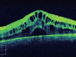
The above picture is an OCT scan of the retina of someone who developed a CRVO. It shows severe swelling of the macula, the central part of the retina. Normal OCT scan of the retina provided below for comparison.

Diagnosis
It is very important to call an ophthalmologist right away if you have any symptoms. He or she will check your eyes thoroughly and talk about treatment options. You may become blind if you do not have treatment for retinal vein occlusion.
During an eye exam, your ophthalmologist will dilate your pupil and look through a special lens at the inside of your eye. Your doctor can diagnose a CRVO or BRVO when examining your eyes based on the appearance of hemorrhages and swelling in the retina. Your doctor may do fluorescein angiography to see what is happening with your retia. Yellow dye (called fluorescein) is injected into a vein, usually in your arm. The dye travels through the blood vessels. A special camera takes photos of the back of your eye as the dye travels throughout its blood vessels. This shows if abnormal new blood vessels are growing under the retina.
Optical coherence tomography (OCT) is another way to look closely at the retina. A machine scans the retina and provides very detailed images of the retina and macula. This helps your doctor find and measure swelling of your macula.
How is retinal vein occlusion treated?
Your ophthalmologist will treat you based on the findings of the exam. However, the main reason for decreased vision from a retinal vein occlusion is swelling in the central part of the retina called macular edema (see picture above). Treatment may include:
Medicine: A drug is given by injections (shots) inside the eye. The medicine helps reduce swelling of the macula. This helps to slow vision loss and in many cases improves vision.
Laser surgery: Your ophthalmologist may use a laser to shrink certain blood vessels in your retina. This is so that they won’t bleed, leak and to prevent them from growing back. Laser surgery may help slow vision loss and perhaps improve vision.
Managing your health: Diabetes, glaucoma, high blood pressure or other health problems can lead to retinal vein occlusion. Taking care of your health can keep you from getting this serious eye problem. A complete medical evaluation is recommended with careful attention to cardiovascular disease. Your ophthalmologist and primary care physician may talk with you about the best ways to manage your health and reduce your risk of retinal vein occlusion from developing in the other eye. There is an 8-10% risk of retinal vein occlusion developing in your other unaffected eye.
Summary
Retinal vein occlusion is when a vein in your retina is blocked. This causes blurry vision or vision loss. It is treated with medication injections, and/or laser surgery and modification (improvement) of health risk factors such as diabetes and high blood pressure.
